Hybrid microscope breakthrough captures molecular movement in 3D – Interesting Engineering
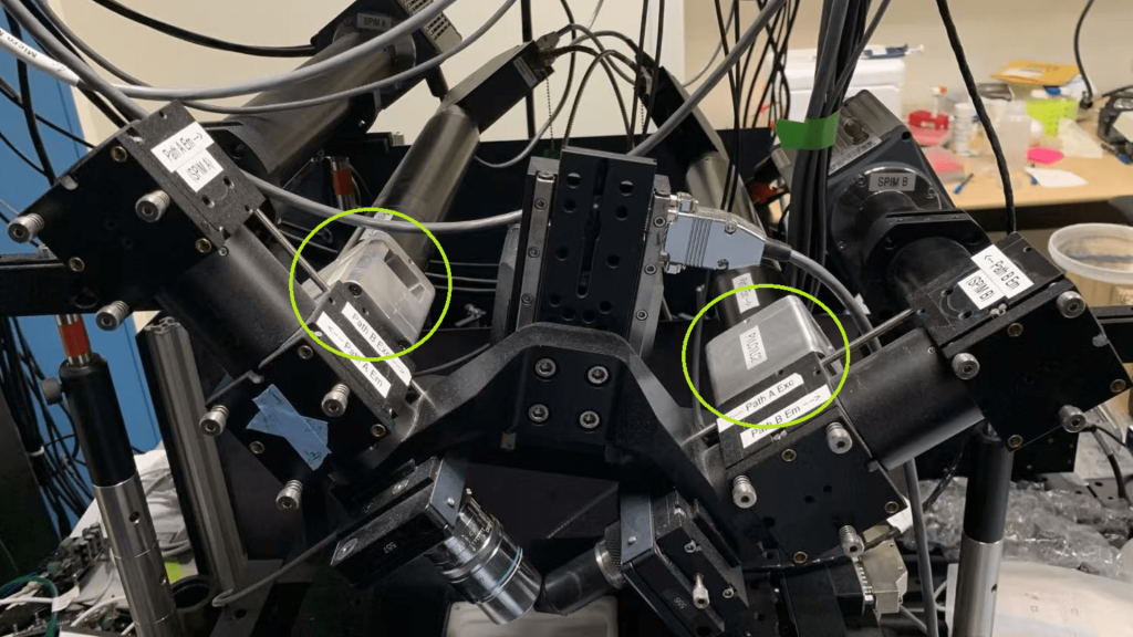
The instrument allows researchers to record changes in 3D protein orientation. a day agoa day ago2 days ago2 days ago2 days ago2 days ago2 days ago2 days ago3 days ago3 days agoan hour ago2 hours ago2 hours ago2 hours ago3 hours ago3 hours ago3 hours ago3 hours ago3 hours ago4 hours agoBojan StojkovskiThe polarized diSPIM, constructed in the Shroff lab at NIH after the idea was conceived at the MBL.Marine Biological Laboratory A hybrid microscope developed at the Marine Biological Laboratory (MBL) allows scientists to capture both the 3D orientation and position of molecular ensembles, such as labeled proteins inside cells.The microscope combines polarized fluorescence, which measures molecular orientation, with a dual-view light sheet microscope (diSPIM) that captures depth details in a sample. This technology can be useful for studying proteins, as they change their 3D orientation in response to their environment to interact with other molecules and perform their functions.According to Talon Chandler of CZ Biohub San Francisco, the study’s first author and a former University of Chicago graduate student who conducted part of this research at MBL, the instrument allows researchers to record changes in 3D protein orientation. This capability provides insights that may be missed when looking only at a molecule’s position. One example is imaging molecules in the spindle of a dividing cell, a challenge that has long been studied at MBL and other research institutions.The study’s co-author, Rudolf Oldenbourg, a senior scientist at MBL, explained that traditional microscopy, including polarized light, can effectively image the spindle when it is perpendicular to the viewing direction. However, when the spindle is tilted, the readout becomes ambiguous. The new instrument overcomes this limitation by adjusting for tilt, allowing researchers to accurately capture both the 3D orientation and position of spindle molecules, such as microtubules.Now, the team aims to improve the system’s speed to capture how the position and orientation of structures change in live samples over time. They also hope that future fluorescent probes will expand its use, allowing researchers to image a wider range of biological structures.The idea for the microscope originated in 2016 through brainstorming sessions among microscopy innovators at MBL. Hari Shroff of HHMI Janelia, then at the National Institutes of Health (NIH) and an MBL Whitman Fellow, was using his custom-built diSPIM microscope at MBL, developed in collaboration with Abhishek Kumar, now at MBL.The diSPIM microscope features two imaging paths that intersect at a right angle, allowing researchers to illuminate and capture the sample from both perspectives. This dual-view approach improves depth resolution compared to a single view and provides greater control over polarization during imaging.Shroff and Oldenbourg recognized that the dual-view microscope could help overcome a limitation of polarized light microscopy – its difficulty in efficiently illuminating a sample with polarized light along the direction of light propagation. By incorporating two orthogonal views, they saw an opportunity to improve the detection of polarized fluorescence and explored using the diSPIM system for such measurements.Shroff collaborated with Patrick La Riviere from the University of Chicago, whose student Talon Chandler joined the project at MBL. Chandler’s doctoral thesis focused on integrating the two systems, working in Oldenbourg’s lab for a year. The team, including Shalin Mehta, outfitted the diSPIM with liquid crystals to control input polarization direction.Chandler dedicated a significant amount of time to exploring how to reconstruct the data and maximize what could be recovered from it. Co-author Min Guo, then at Shroff’s previous lab at NIH, also worked extensively on this aspect, and together, they achieved their goal of full 3D reconstructions of molecular orientation and position.Bojan Stojkovski Bojan Stojkovski is a freelance journalist based in Skopje, North Macedonia, covering foreign policy and technology for more than a decade. His work has appeared in Foreign Policy, ZDNet, and Nature.Stay up-to-date on engineering, tech, space, and science news with The Blueprint.By clicking sign up, you confirm that you accept this site’s Terms of Use and Privacy Policy21 hours ago21 hours agoa day agoa day agoPremiumIE PROFollow
Source: https://interestingengineering.com/science/hybrid-microscope-captures-molecular-movement-3d